 Movie
Movie Controller
 Structure viewers
 About EMN search


-Search query
-Search result
Showing all 43 items for (author: chlanda & p)
EMDB-15964: 
Subtomogram averaging of SARS-CoV-2 nsp3-4 delta Ubl1-Mac1 obtained from cryo-ET of cryo-FIB milled VeroE6 cells transfected with nsp3-4 delta Ubl1-Mac1
Method: subtomogram averaging / : Chlanda P, Zimmermann L
EMDB-15965:
Subtomogram average of SARS-CoV-2 nsp3-4 delate Ubl1-Ubl2 obtained from cryo-ET of cryo-FIB milled VeroE6 cells transfected with nsp3-4 delta Ubl1-Ubl2
Method: subtomogram averaging / : Chlanda P, Zimmermann L
EMDB-15963:
Subtomogram average SARS-CoV-2 nsp3-4 from cryo-ET of VeroE6 expressing nsp3-4 (filtered to 20A resolution).
Method: subtomogram averaging / : Chlanda P, Zimmermann L

EMDB-15925:
Cryo-electron tomogram acquired on cryo-lamella of VeroE6 cells transfected with SARS-CoV-2 HA-nsp3-4-V5, plunge frozen at 16 hpt
Method: electron tomography / : Zimmermann L, Chlanda P
EMDB-15926:
Cryo-electron tomogram of VeroE6 cells transfected with HA-nsp3-deltaUbl1-Ubl2-nsp4-V5, plunge frozen at 16 hpt
Method: electron tomography / : Zimmermann L, Chlanda P
EMDB-15927:
Cryo-electron tomogram of VeroE6 cells transfected with HA-nsp3-deltaUbl1-Mac1-nsp4-V5. Cells were plunge-frozen at 16h post-transfection and subjected to cryo-FIB milling.
Method: electron tomography / : Chlanda P, Zimmermann L
EMDB-15928:
Cryo-electron tomogram of VeroE6 cells transfected with HA-GG>AA-nsp4-V5, plunge-frozen at 16 hpt and subjected to cryo-FIB milling.
Method: electron tomography / : Chlanda P, Zimmermann L
EMDB-15929:
Cryo-electron tomogram of VeroE6 cells transfected with nsp3-4, vitrified by plunge-freezing at 16 hours post transfection and processed by cryo-FIB milling.
Method: electron tomography / : Chlanda P, Zimmermann L
EMDB-15244:
Tomogram of an Ebola VLP composed of GP, VP40, NP, VP24 and VP35 at pH 7.4 (Figure 1A-D)
Method: electron tomography / : Winter SL, Chlanda P
EMDB-15268:
Tomogram of an Ebola VLP composed of VP40 at pH 4.5 (Figure 1J)
Method: electron tomography / : Winter SL, Chlanda P
EMDB-15951:
Tomogram of an EBOV-infected Huh7 cell showing a late endosome with internalized EBOV particles
Method: electron tomography / : Winter SL, Chlanda P
EMDB-15956:
Tomogram of an extracellular EBOV particle adjacent to an EBOV-infected Huh7 cell
Method: electron tomography / : Winter SL, Chlanda P
EMDB-16128:
Tomogram of a late endosome of A549 cell infected with influenza A virus.
Method: electron tomography / : Klein S, Chlanda P
EMDB-16129:
Tomogram of a late endosome of A549 cell infected with influenza A virus (Figure 6C).
Method: electron tomography / : Klein S, Chlanda P

EMDB-16130:
Tomogram of a late endosome of A549 cell infected with influenza A virus (Figure 6E)
Method: electron tomography / : Klein S, Chlanda P
EMDB-16131:
Tomogram of a late endosome of A549 cell infected with influenza A virus (Figure 6G)
Method: electron tomography / : Klein S, Chlanda P
EMDB-16132:
Tomogram of a late endosome of A549 cell infected with influenza A virus (Figure 6I,K,M)
Method: electron tomography / : Klein S, Chlanda P
EMDB-16133:
Tomogram of a late endosome of A549 cell infected with influenza A virus (Figure 6O)
Method: electron tomography / : Klein S, Chlanda P
EMDB-15705:
Tomogram of a late endosome of A549 cell (Figure 1R)
Method: electron tomography / : Klein S, Chlanda P
EMDB-15707:
Tomogram of a late endosome of A549 cell treated with IFN-beta (Figure 1T)
Method: electron tomography / : Klein S, Chlanda P
EMDB-15708:
Tomogram of a late endosome of A549-IFITM3 cell (Figure 1V)
Method: electron tomography / : Klein S, Chlanda P
EMDB-15131:
Tomogram of a late endosome of A549-IFITM3 cells infected with influenza A virus (Figure S7)
Method: electron tomography / : Klein S, Chlanda P
EMDB-15130:
Tomogram of a late endosome of A549-IFITM3 cells infected with influenza A virus (Figure 4E)
Method: electron tomography / : Klein S, Chlanda P
EMDB-15132:
Tomogram of a late endosome of A549-IFITM3 cells infected with influenza A virus (Figure S8)
Method: electron tomography / : Klein S, Chlanda P

EMDB-15133:
Tomogram of a late endosome of A549-IFITM3 cells infected with influenza A virus (Figure S9)
Method: electron tomography / : Klein S, Chlanda P

EMDB-10819:
Tomogram of Lamellar body in A549 cell
Method: electron tomography / : Klein S, Wimmer B, Winter S, Kolovou A, Laketa V, Chlanda P

EMDB-10820:
Tomogram of lamellar body (A549 cell expressing ABCA3-eGFP)
Method: electron tomography / : Klein S, Wimmer B, Winter S, Kolovou A, Laketa W, Chlanda P

EMDB-10818:
Tomogram of Lamellar body in A549 cell expressing ABCA3-eGFP
Method: electron tomography / : Klein S, Wimmer B, Winter S, Kolovou A, Laketa V, Chlanda P

EMDB-11929:
In situ cryo-ET of HSAEpC primary lung cells
Method: electron tomography / : Klein S, Wimmer B, Winter S, Kolovou A, Laketa V, Chlanda P
EMDB-11865:
Tomogram of SARS-CoV-2 infected and cryo-FIB milled VeroE6 cells
Method: electron tomography / : Klein S, Cortese M, Winter SL, Wachsmuth-Melm M, Neufeldt CJ, Cerikan B, Stanifer ML, Boulant S, Bartenschlager R, Chlanda P

EMDB-11931:
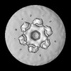
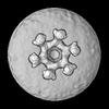
Subtomogram average of outer membrane dome protein (OMDP)-C8 symmetrized
Method: subtomogram averaging / : Klein S, Wimmer B, Winter S, Kolovou A, Laketa V, Chlanda P
EMDB-11863:
Cryo-ET of SARS-CoV-2 infected VeroE6 cells showing budding virions.
Method: electron tomography / : Klein S, Cortese M, Winter SL, Wachsmuth-Melm M, Neufeldt CJ, Cerikan B, Stanifer ML, Boulant S, Bartenschlager R, Chlanda P
EMDB-11866:
SARS-CoV-2 structure and replication characterized by in situ cryo-electron tomography
Method: electron tomography / : Klein S, Cortese M, Winter SL, Wachsmuth-Melm M, Neufleldt CJ, Cerikan B, Stanifer ML, Boulant S, Bartenschlager R, Chlanda P
EMDB-11867:
Cryo-ET of SARS-CoV-2 virions released from VerE6 cells
Method: electron tomography / : Klein S, Cortese M, Winter SL, Wachsmuth-Melm M, Neufeldt CJ, Cerikan B, Stanifer ML, Boulant S, Bartenschlager R, Chlanda P
EMDB-11868:
Subtomogram average of SARS-CoV-2 vRNP
Method: subtomogram averaging / : Klein S, Cortese M, Winter SL, Wachsmuth-Melm M, Neufeldt CJ, Cerikan B, Stanifer ML, Boulant S, Bartenschlager R, Chlanda P

EMDB-8843:
Influenza A virus-like particles containing HA-MAY, NA, M1, and M2 proteins (MAY denotes three amino acids that replace three cysteine residues in the cytoplasmic tail of hemagglutinin)
Method: electron tomography / : Chlanda P, Zimmerberg J

EMDB-8844:
Influenza A virus-like particles containing Hemagglutinin, Neuraminidase, M1, and M2 proteins
Method: electron tomography / : Chlanda P, Zimmerberg J

EMDB-8845:
Influenza A virus-like particles containing Hemagglutinin, Neuraminidase, M1, and M2 proteins
Method: electron tomography / : Chlanda P, Zimmerberg J

EMDB-8846:
Influenza A virus-like particles containing HA-MAY, NA, M1, and M2 proteins incubated with liposomes at pH 5 (MAY denotes three amino acids that replace three cysteines in the cytoplasmic tail of hemagglutinin)
Method: electron tomography / : Chlanda P, Zimmerberg J

EMDB-8088:
G1S VLP_noVPP
Method: electron tomography / : Chlanda P, Zimmerberg J

EMDB-8089:
G1S VLP_VPP
Method: electron tomography / : Chlanda P, Zimmerberg J

EMDB-2103:
Heritable yeast prions have a highly organized 3-dimensional architecture with inter-fiber structures
Method: subtomogram averaging / : Frangakis A, Saibil HR

EMDB-2104:
Heritable yeast prions have a highly organized 3-dimensional architecture with inter-fiber structures
Method: subtomogram averaging / : Frangakis A, Saibil HR
 wwPDB to switch to version 3 of the EMDB data model
